Wanderröte Verwechseln refers to Confusing Erythema Migrans. Erythema migrans (EM) is a prototypical skin illness of Lyme disease, an Erkrankung that is infectious because of a tick (zecken) or spirochete, Borrelia burgdorferi, and other species of spirochete in the United States, aus across parts of Europe in the Asian continent. Referred to as an increasing, erythematous rash, and capable of taking the form of a bull-eye, EM is a typical community sign of Lyme disease, which develops between 3 and 30 days after einem ersten Angriff by a tagen. Due to its inconsistent nature and similar appearance to some of the other skin conditions, it is commonly misdiagnosed. Die Ursachen for this misdiagnosis can be attributed to its resemblance to other Erkrankungen, no matter how it appears, was eine gewisse Verwirrung nach Jahren verursachen kann und dauert Wochen nach dem ersten Stich.
Table of Contents
Characteristics of Erythema Migrans
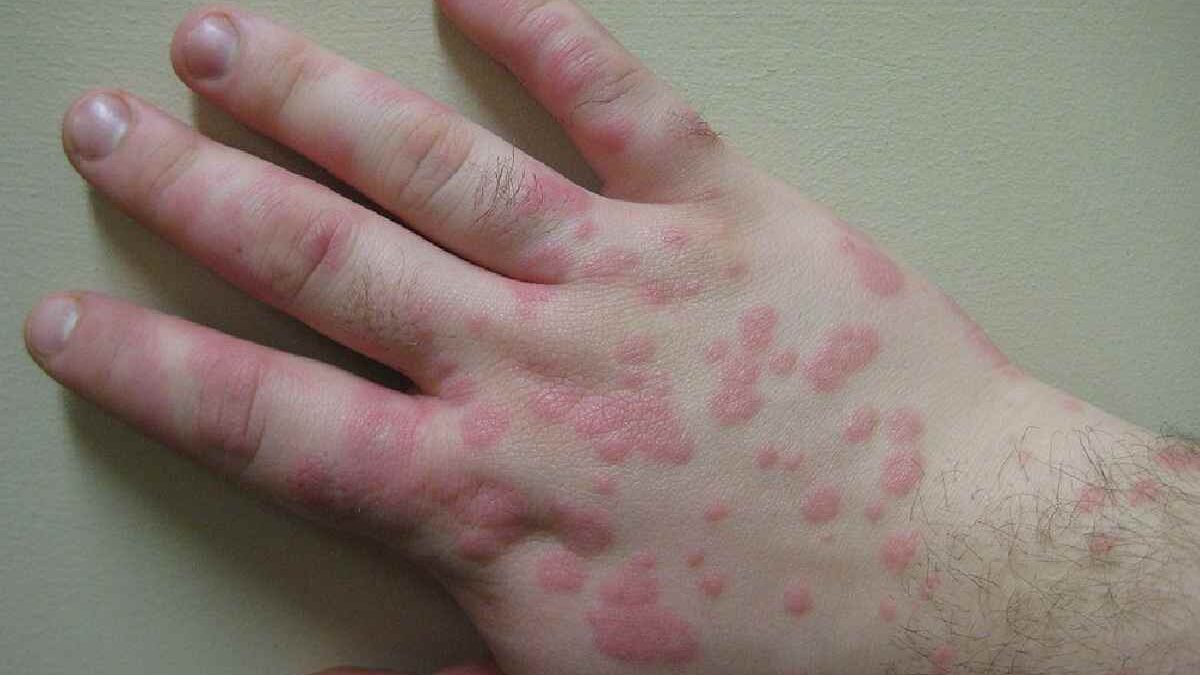
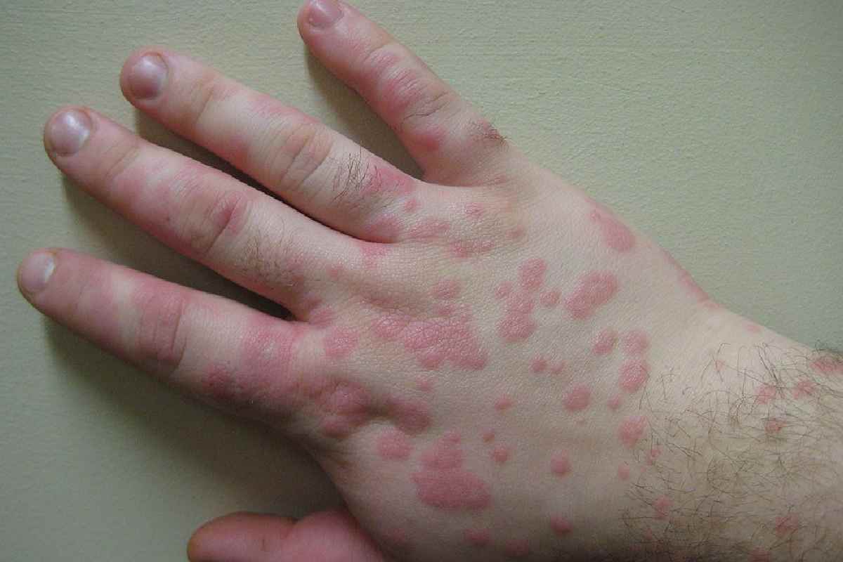
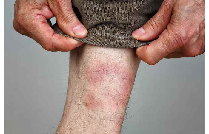
EM usually goes through some stages, and initially there is a small red macule or papule at the site of a zecke bite in Deutschland, and then it grows over weeks. This occurs in the form of a circular or oval rash with a central clearing that provides the bull’s-eye look, but only 20-30 percent of cases have the pattern. The size of the rash can be as big as 70 cm or as little as 5 cm, and it is often asymptomatic except that some patients complain of mild itchiness, burning, or akute schmerzen. EM may bring with it other systemic symptoms; in disseminated Lyme disease, especially during the early phase, fever, fatigue, kopfschmerzen, and arthralgia are common, and when the Nervensystem is affected, it can lead to more severe complications such as neuroborreliose or require treatment with antibiotika some weeks to monate after the initial zeckenstich.
EM is variable, making it difficult to identify. The unusual features are non-homogeneous erythema or multiple lesions, or irregular in shape. The rash can be less erythematous, more violaceous, or hyperpigmented on dark skin tones to further confound diagnosis.
Conditions Mimicking Erythema Migrans
Certain dermatological problems are similar to EM, and this leads to confusion in the diagnosis. These include:
- Allergic contact dermatitis: The contact dermatitis occurs after an individual is exposed to allergens such as poison ivy or metals and manifests in erythematous, pruritic eruptions. As opposed to EM, it usually has vesicles or scaling and exhibits a pattern of allergen exposure, e.g., linear streaks.
- Tinea Corporis (Ringworm): Tinea corporis appears as ring-shaped, scaly lesions with raised edges that resemble the bull’s-eye picture of EM. Tinea corporis is, however, more pronounced in its scale and responds to antifungal medication, hence, unlike EM.
- Urticaria: Hives: transient, erythematous wheals which sometimes merge to form bigger plaques. Urticaria is characterized as extremely pruritic, changes hour to hour, and is commonly related to allergic methods as compared to EM.
- Fixed Drug Eruption: Some drugs trigger a recurrent, well-defined erythematous eruption at certain sites. These lesions can be similar to EM but are usually drug-related and disappear once the causative agent is removed.
- Bacterial skin infections: Bacterial skin infections, such as cellulitis, present with diffuse erythema, warmth, and tenderness, which may be confused with homogeneous EM. The disease cellulitis is faster and more likely to be accompanied by lymphangitis and fever as opposed to most cases of EM.
- Granuloma Annulare: This benign condition presents as annular, asymptomatic plaques with a central clearing, resembling EM. It lacks systemic symptoms and is more chronic, often persisting for months.
Diagnostic Challenges
The clinical variation and co-occurrence of EM are the reasons that it is misdiagnosed. The main issues are:
Absence of Patient History: Patients will not remember having been bitten by a tick since only 50-70 percent of them do. Lack of this history makes it harder to associate this rash with Lyme disease.
Non-classic presentations: Multiple lesions or homogenous erythema, which occur in non-classic EM, can be confused with other dermatoses, impeding the establishment of Lyme disease.
Geographic Variability: The presence of Lyme disease is dependent on the geographic location, which implies a clinician’s suspicion. EM can also be disregarded in non-endemic regions in favor of other, more widespread conditions.
Serological weaknesses: Serologic screening tests of early Lyme disease are often negative because the antibodies take 2-6 weeks to develop. During the EM stage, the use of serology may cause a false negative.
Physician Awareness: The primary care community physicians might have no exposure to the variety of manifestations of EM, especially in parts of the country where there are low cases of Lyme disease.
Importance of Accurate Diagnosis
Accurate diagnosis of EM is important when initiating timely Behandlung and preventing the progression of Lyme krankheit in kindern. Early Lyme disease can lead to chronische Borreliose but can be successfully cured using oral antibiotics (e.g., doxycycline or doxycyclin, amoxicillin) during 10-21 days, eliminating EM and the development of such complications as arthritis, carditis, or the emergence of neurological implications and verschiedenen beschwerden. The risk of misdiagnosis, however, may result in delayed management and the need for antibiotische therapie, thus raising the risk of the disseminated disease or ineffective interventions, including the use of antifungals to treat tinea corporis, which does not work with Lyme disease. Additionally, a proper st diagnosis of Lyme disease is critical to avoid these complications.
Furthermore, EM can be wrongly treated as a benign disease, such as granuloma annulare, potentially delaying the exploration of the systemic complaints, whereas the diagnosis of cellulitis can lead to an unevoked antibiotics scheme, causing the eventual development of resistance. Proper diagnosis is also a matter of public health because Lyme disease is reportable in many areas, which can contribute to surveillance and prevention activities.
Strategies to Improve Diagnosis
To lessen the misunderstanding of EM, it is possible to adopt some strategies:
Clinical Education: The education of healthcare personnel on the various manifestations of EM, such as unusual varieties and manifestations in various skin tones, ensures that the healthcare provider performs the diagnosis more accurately. In the non-endemic and endemic regions, CME activities must focus on Lyme disease.
Awareness of Patients: Community health and education efforts can help them know how to avoid tick bites and the need to observe the development of rashes and seek medical attention promptly.
Diagnostic Algorithms: The integration of clinical decision elements to include patient histories, specifics of the rash, and the regional presence of Lyme disease can facilitate the providers in making accurate diagnoses.
Dermoscopy and Imaging; Non-invasive techniques, e.g., dermoscopy, can demonstrate minute detail, including peripheral erythema or central clearing, which helps to distinguish between EM and fac-similes. State-of-the-art imaging, which is rarely necessary, can aid in solving such mysterious cases.
Molecular Testing. In the case of the uncertain situation, Borrelia DNA testing of skin biopsies by polymerase chain reaction can confirm Lyme disease when serology is unhelpful.
Conclusion
Erythema migrans would be a key indicator of Lyme disease, but it appears differently and is similar to other skin diseases or conditions, thus creating a big dilemma. This may result in delays of the treatment process and aggravate patients, on one hand, and deplete healthcare resources, on the other hand. Incorporation of learned clues about the characteristics of EM, identifying mimics, and managing diagnostic barriers will help clinicians to identify early Lyme disease and manage it.